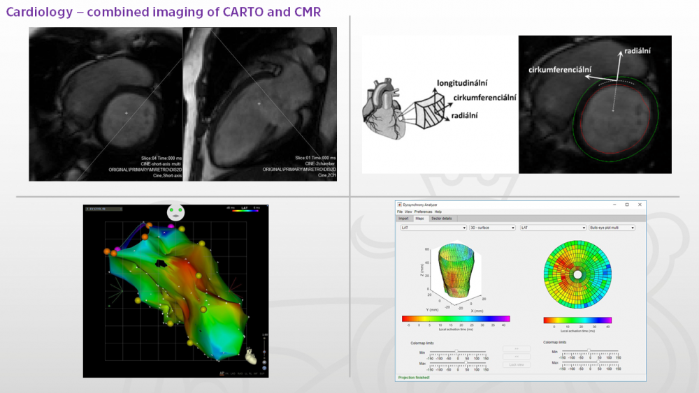
- Imaging methods for detecting symptoms of electromechanical cardiac dyssynchrony
-
Scientific team: Clinical applications of imaging systems and methods Contact person: doc. Ing. Jiří Hozman, Ph.D., +420 728 335 738 Description of the activities:
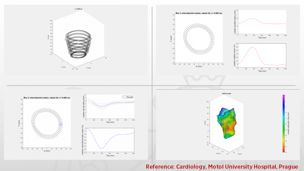
The field of electrical dyssynchrony of the heart is very well methodologically supported and the existing criteria and experience are sufficient to address the patients' problems. However, no one has yet systematically addressed the combination of cardiac MRI (CMR) and left ventricular electroanatomical mapping (CARTO) data, which could be used as a suitable method to refine the nature of cardiac electromechanical dyssynchrony and thus to improve the effectiveness of treatment of chronic heart failure. It was possible to design a method for calculation and display of electrical and mechanical parameters, which were implemented in an application usable in clinical practice. Subsequently, it was statistically verified that the values of the mechanical parameters, i.e. maximum radial strain rate, minimum circumferential strain rate and the electrical parameter local activation time, differed significantly between the patient set and the healthy control set. These parameters were then determined to be suitable for assessing the presence of electromechanical dyssynchrony.
Competencies and infrastructure:
Modeling and simulation processes applied onto the medical imaging modalities, analysis and synthesis within the medical imaging systems and methods. Matlab and Segment SW for strain analysis and fusion or merge of the results from CMR and from the CARTO. Dyssynchrony Analyser as app in Matlab.
Other info:
- Cooperation
- Clinic of Cardiology Motol University Hospital, Assoc. Prof. MUDr. Lucie Riedlbauchová, Ph.D.
- Publications
- Riedlbauchova, L.; Lozek, M.; Adla, T.; Suchanek, V.; Huptych, M.; Hozman, J. et al. Role of internal stretch fraction parameter in quantification of mechanical dyssynchrony and prediction of cardiac resynchronization therapy outcome European Heart Journal. 2019, 40(Issue Supplement_1), 3556. ISSN 0195-668X.
- Riedlbauchová, L.; Adla, T.; Suchánek, V.; Ložek, M.; Tomis, J.; Hozman, J.; Tomek, V.; Veselka, J. et al. Is left bundle branch block pattern on the ECG caused by variable ventricular activation sequence? PACE: Pacing and Clinical Electrophysiology. 2020, 43(5), 486-494. ISSN 0147-8389.
- Riedlbauchova, L.; Adla, T.; Suchanek, V.; Lozek, M.; Tomis, J.; Hozman, J.; Tomek, V.; Veselka, J. et al. Is the typical left bundle branch block pattern on the ECG caused by variable ventricular activation sequences? European Journal of Heart Failure. 2018, 20(Suppl. S1), 34-35. ISSN 1388-9842.
- Adla, T.; Suchánek, V.; Riedlbauchová, L.; Ložek, M.; Tomis, J.; Hozman, J.; Janoušek, J.; Roček, M. Comparison of Two Methods of Left Ventricular Global Strain Assessment in Normal Subjects: Feature Tracking Versus Tagging European Heart Journal - Cardiovascular Imaging. 2017, 18(2), ii165. ISSN 2047-2404.
- Projects
- Cooperation
- Anatomically faithful patient heart models for planning mechanical left atrial appendage occlusion
-
Scientific team: Bio-Electromagnetism Contact person: Assoc. Prof. Ing. Ondřej Fišer, Ph.D. Description of the activities:
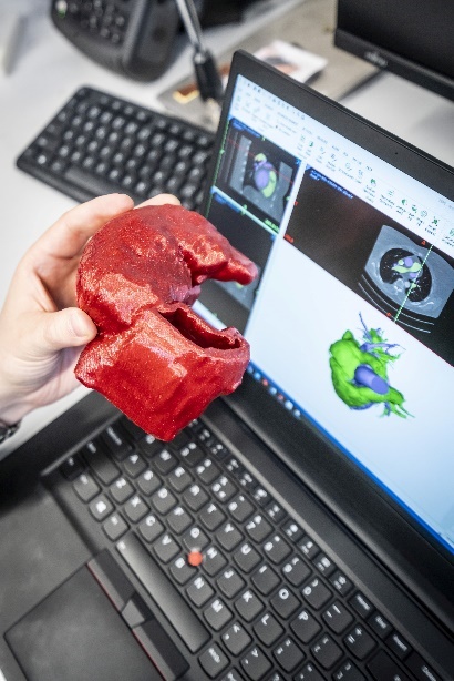
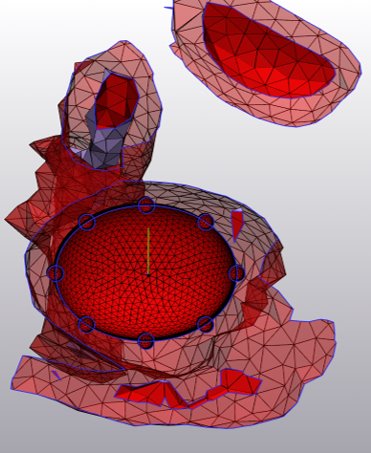
Assoc. prof. Ondřej Fišer, Dr. Tomáš Pokorný, and Prof. David Vrba from the Bioelectromagnetism research team are collaborating with doctors from the Cardiology Clinic of the Královské Vinohrady University Hospital (FNKV) and the Karlovy Vary Regional Hospital to increase the success and safety of catheter closure of the left atrial appendage. The procedure is indicated for patients suffering from atrial fibrillation with an increased risk of thrombus formation, in whom anticoagulant therapy fails due to the high risk of internal bleeding. These patients are at significant risk of stroke, as thrombus most often occurs in the left atrial appendage, from where it most often travels through the left ventricle directly to the brain. Closing the left atrial appendage prevents thrombus formation and thus reduces the risk of stroke. Due to the high morphological variability of the left atrial appendages, which is individual for each patient, it is important to know the ideal location for puncturing the interatrial septum to ensure its successful closure. For this purpose, an individual 3D computer model of the patient's heart is created at FBMI before each procedure. A template is first created from the patient's CT images, which is then used to print a real model on a 3D printer.
Thanks to these models, doctors at the FNKV Cardiology Clinic are able to find the ideal location for puncturing the interatrial septum, which is one of the key moments of the procedure that can significantly affect its further course. Thanks to the possibility of “in vitro” simulation, the procedure can be individualized with regard to the anatomical conditions of the patient's heart, which is important not only for the effectiveness but also for the safety of the procedure. The original collaboration between the Bioelectromagnetism team, the Cardiology Clinic at FNKV, and the Karlovy Vary Regional Hospital has gradually grown into a multicenter study currently involving six centers and has helped optimize left atrial appendage closure for more than 50 patients with atrial fibrillation.
Competencies and infrastructure:
- Preparation of anatomically faithful heart models from patient CT/MRI images. 3D printing of anatomical models from rigid and flexible materials.
- Materialise Mimics, Průša SL1 and i3 MK3 3D printers
Other info:
- Cooperation
- Kardiologická klinika FNKV, Karlovarská krajská nemocnice, …
- Projects
- Horizon 2020 - Fast Track to Innovation, „Left Atrial Appendage Electrical Isolation via Bio-photonic Optical Confirmation to Treat Persistent Atrial Fibrillation- LAA-START“, Project No. 831117
- Design of applicators and numerical modelling of radiofrequency and microwave ablation and electroporation
-
Scientific team: Bio-Electromagnetism Contact person: Prof. Ing. David Vrba, Ph.D. Description of the activities:
An international consortium of companies and universities from three countries, including the Faculty of Biomedical Engineering at the Czech Technical University in Prague, received a three-year grant in 2019 from the European Commission's prestigious Horizon 2020 program, Fast Track to Innovation, worth more than one million euros to complete the development of a system that will reduce the risk of stroke and heart failure in patients with atrial fibrillation, i.e., irregular heartbeat caused by abnormal electrical activity in the heart.
Irish company AuriGen Medical designed the concept and developed a unique prototype of a minimally invasive cardiac implant that permanently electrically and mechanically isolates the left atrial appendage in a single procedure. This is a very common site of blood clots in patients with chronic atrial fibrillation, which can cause strokes or heart failure. There are approximately more than 10 million patients in the EU and the US.
The aim of the grant Left Atrial Appendage Electrical Isolation via Bio-photonic Optical Confirmation to Treat Persistent Atrial Fibrillation was to complete the development of this system and subsequently introduce it into practice, thereby minimizing the formation of blood clots and thus fatal damage to the patient's brain and heart.
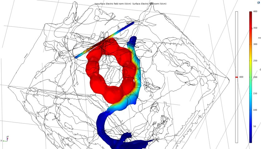
As part of this project, Prof. David Vrba, Assoc. Prof. Ondřej Fišer, and Prof. Jan Vrba from the Bioelectromagnetism team at the Department of Biomedical Technology, FBME, participated in the design and optimization of the geometry of the applicator, which will be responsible for radiofrequency ablation of cardiac arrhythmia within the developed system.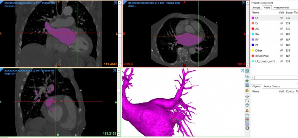
Competencies and infrastructure:
- Numerical modeling of electric fields, currents, and induced heating in biological tissues during ablation. Calculations to evaluate the success and extent of ablation and the risk of unwanted thermal damage to adjacent tissues. Design and optimization of applicator geometry and methods. Preparation of anatomically accurate heart models from patient CT/MRI images.
- Sairem 200W, 2.45 GHz semiconductor microwave generator. COMSOL Multiphysics, Sim4Life, and Materialise Mimics segmentation software numerical simulators.
Other info:
- Cooperation
- AuriGen Medical, Ireland
- Projects
- Horizon 2020 - Fast Track to Innovation, „Left Atrial Appendage Electrical Isolation via Bio-photonic Optical Confirmation to Treat Persistent Atrial Fibrillation- LAA-START“, Project No. 831117
- Development and production of hardware and software devices for recording, visualisation and analysis of ECG signals
-
Scientific team: Team of Biomechanics and Assistive Technology Contact person: Assoc. Prof. Ing. Patrik Kutilek, M.Sc., Ph.D., +420 728 335 738 Description of the activities:
We are developing a wearable multisensory system that enables real-time recording of ECG, respiratory curves, and bioimpedance. Since the device has been tested in high-pressure and high-humidity environments, it can be used in very specific conditions.
In terms of software, we are developing modular applications for recording, visualizing, and analyzing physiological data. We focus primarily on methods for detecting increased cognitive load using analytical methods that evaluate heart rate variability (e.g., HRV or nonlinear analysis) and artificial intelligence methods. The methods are developed in collaboration with institutions engaged in research primarily in the field of psychology (NÚDZ, ÚPOL, INESAN).
We carry out development and research within the framework of project activities (TA ČR, GA ČR), contract research, and student work.Competencies and infrastructure:
Other info:
- Cooperation
- Projects
- Application of BSPM in patients with resynchronization therapy
-
Scientific team: Contact person: Assoc. Prof. Mgr. Ksenia Sedova, Ph.D., +420 775 535 282 Description of the activities:
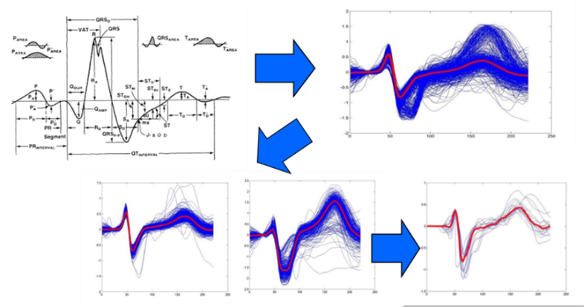
The concept of the transdisciplinary project is based on research collaboration with the IKEM Cardiology Clinic (Prof. Josef Kautzner, MD, PhD, FESC). In cooperation with the Department of Cardiology, University Medical Center Utrecht (Utrecht, Netherlands), computer simulation of myocardial electrophysiological processes in the heart and application of inverse problem methods for patient-specific visualization of ventricular activity and torso reconstruction are being performed (Peter Van Dam, Ph.D.). Colleagues from the Polytechnic University (Università Politecnica della Marche, Ancona, Italy) are working on the analysis of electrocardiographic signals of the heart as part of this project. They are responsible for the design of Point2ECG, a new interactive application for the automatic identification of ECG reference points, enabling their visual inspection and correction (Agnese Sbrollini, Ph. D., Prof. Laura Burattini, Ph.D.).
Competencies and infrastructure:
Other info:
- Cooperation
- prof. MUDr. Josef Kautzner, CSc., FESC (Department of Cardiology, Institute for Clinical and Experimental Medicine, Prague, Czech Republic)
- Peter van Dam, Ph.D. (University Medical Center Utrecht, Utrecht, The Netherlands; ECG Excellence BV, Nieuwerbrug, The Netherlands)
- Agnese Sbrollini, Ph.D. (Università Politecnica della Marche, Ancona, Italy) prof. Laura Burattini, Ph.D. (Università Politecnica della Marche, Ancona, Italy)
- Publications
- Sedova K, Repin K, Donin G, Van Dam P, Kautzner J. Clinical utility of body surface potential mapping in CRT patients. Arrhythm Electrophysiol Rev 2021;10(2):113-119. JCR Q1 (Scopus).
- Projects
- Cooperation
- Prediction of ventricular fibrillation in patients with acute myocardial infarction
-
Scientific team: Contact person: Assoc. Prof. Mgr. Ksenia Sedova, Ph.D., +420 775 535 282 Description of the activities:
The subject of this clinical research is to address two main issues: 1. finding electrocardiographic parameters of depolarization and repolarization associated with the onset of ventricular fibrillation in patients with acute coronary syndrome, 2. developing an algorithm for automated determination of these parameters.
Competencies and infrastructure:
Other info:
- Cooperation
- prof. Pyotr Platonov, M.D., Ph.D. (Department of Cardiology, Clinical Sciences, Lund University, Lund, Sweden)
- Marina Demidova, M.D., Ph.D. (Department of Cardiology, Clinical Sciences, Lund University, Lund, Sweden;
- V.A. Almazov National Medical Research Center, Saint Petersburg, Russia)
- Publications
- Sedova KA, Demidova MM, Azarov JE, Hejda J, Carlson J, Bernikova OG, Arteyeva N, Erlinge D, Platonov PG. Terminal T-wave inversion predicts reperfusion tachyarrhythmias in STEMI. J Electrocardiol. 2022; 71:28-31. doi: 10.1016/j.jelectrocard.2021.12.008.
- Projects
- Cooperation
- Mechanisms of ventricular fibrillation and arrhythmogenic substrate modification
-
Scientific team: Contact person: Assoc. Prof. Mgr. Ksenia Sedova, Ph.D., +420 775 535 282 Description of the activities:
The main challenge of basic research is to determine the role of various electrophysiological abnormalities in the onset of ventricular fibrillation, followed by the development of a new tool for classifying the risk of ventricular fibrillation based on ECG analysis
Competencies and infrastructure:
Other info:
- Cooperation
- Jan Azarov, Ph.D., Olesya Bernikova, M.D., Ph.D., Alexandra Durkina, Department of Cardiac Physiology, Institute of Physiology, Komi Science Center, Ural Branch, Russian Academy of Sciences, Syktyvkar, Russia.
- Publications
- Bernikova OG, Durkina AV, Sedova KA, Azarov JE. Determinants of reperfusion arrhythmias: action potential duration versus dispersion of repolarization. J Physiol Pharmacol. 2021; 72(5). doi: 10.26402/jpp.2021.5.04
- Durkina AV, Bernikova OG, Mikhaleva NJ, Paderin NM, Sedova KA, Gonotkov MA, Kuzmin VS, Azarov JE. Melatonin pretreatment does not modify extrasystolic burden in the rat ischemia-reperfusion model. J Physiol Pharmacol. 2021;72(1). doi: 10.26402/jpp.2021.1.15
- Bernikova OG, Vaykshnorayte MA, Ovechkin AO, Sedova KA, Kharin SN, Azarov JE. Preventive Administration of Melatonin Attenuates Electrophysiological Consequences of Myocardial Ischemia. Bull Exp Biol Med. 2020;169(3):328-331. doi: 10.1007/s10517-020-04881-y
- Bernikova OG, Sedova KA, Durkina AV, Azarov JE. Managing of ventricular reperfusion tachyarrhythmias - focus on a perfused myocardium. J Physiol Pharmacol. 2019;70(5). doi: 10.26402/jpp.2019.5.11.
- Projects
- Cooperation
- Measurement, processing and analysis of multi-lead electrocardiography (ECG) recordings (= Body Surface Potential Mapping)
-
Scientific team: Contact person: Description of the activities:
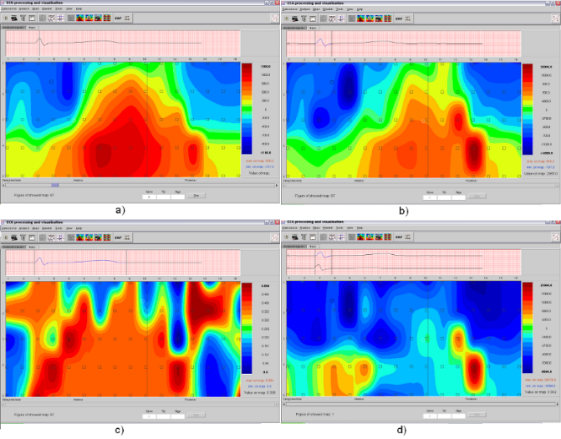
The entire surface potential processing chain has been created. An application has been developed for their pre-processing, which allows for the resolution of complex interference patterns arising from non-ideal measurement settings. An application has been developed for mapping surface potentials, which is the standard method for visualizing these potentials. The application offers basic mapping methods that enable further processing and evaluation of records.
This task was primarily addressed within the project Characteristics of electromechanical dyssynchrony predicting the effect of cardiac resynchronization therapy (CRT)Artificial intelligence methods for detecting pathological ECG patterns (ambulatory 12-lead, Holter)
HRV analysis from wearable electronics
PPG curve analysisExample solution: diagnosis of ventricular tachycardia, fibrillation, and flutter from an electrocardiogram. These heart rhythm disorders are among the most life-threatening arrhythmias. An automatic detection method was developed for the Holter ECG monitoring system. The proposed algorithm is based on detection in the spectral domain supported by detection in the time domain. The result of the work was verified by discriminating the detected arrhythmias from normal ECG recordings and by discriminating them from noise.
Competencies and infrastructure:
Other info:
- Cooperation
- Projects
- Development of recellularized cardiovascular replacements and patches based on decellularized and nanofibrous carriers using tissue engineering methods, including 3D bioprinting and culturing in automated bioreactors
-
Scientific team: Bioreactors for tissue and organ replacements Contact person: Description of the activities:
Development of recellularized cardiovascular grafts and patches based on decellularized and nanofiber carriers using tissue engineering methods, including 3D bioprinting and cultivation in automated bioreactors. Tissue decellularization is performed in specially developed automated systems ensuring homogeneous decellularization of tissue (venous, arterial grafts, and pericardium, human and animal). To reduce immunogenicity and thrombogenicity and increase the overall remodeling capacity of such implants in vivo, the substrates are recellularized using stromal cells from adipose tissue or stem cells from umbilical cord blood and precultured in specialized bioreactors. These special systems simulate the physiological conditions of the vascular wall in vitro and promote the proliferation of cell culture and its differentiation towards the smooth muscle and endothelial phenotype. To ensure the homogeneity of the recellularization of the prepared carriers, a 3D bioprinting method using collagen bioinks is used. The prepared implantable patches and replacements are tested in an animal model – the carotid basin – for 1 and 6 months. The patency of the implants is tested by angiographic imaging and subsequent histopathological analysis in terms of implant integration, overall remodeling and re-epithelialization, patency, and intimal hyperplasia formation. The technology for the preparation of decellularized tissues and culture systems is optimized in terms of compliance with GLP/GMP regulatory requirements in collaboration with a commercial entity.
Competencies and infrastructure:
Other info:
- Cooperation
- Projects
- Research in the field of telemonitoring of cardiovascular diseases
-
Scientific team: Telemedicine and diabetes Contact person: Description of the activities:
The Telemonitoring and Diabetes research team has long focused on the use of telemonitoring (mobile applications, wearable electronics) in the diagnosis and treatment of the most common chronic cardiovascular diseases, primarily arterial hypertension and chronic heart failure.
The team created a telemedicine system focused on cardiovascular diseases and conducted a clinical study on it in 2019-21 involving 150 patients who were monitored for at least 3 months. The aim of the study was to compare the modified methodology of home blood pressure (BP) monitoring with several other methods of BP measurement in the doctor's office. The modification of home monitoring consisted of supplementing BP measurement with monitoring of physical activity, especially before the actual BP measurement. Some results have already been published (e.g., https://ep.liu.se/en/conference-article.aspx?series=ecp&issue=161&Article_No=28 ), while the main results are still being prepared for publication. Another clinical study focused on the use of telemonitoring in patients with chronic heart failure is currently being prepared.
The team has technologies for long-term monitoring of physical activity, heart rate, blood pressure, SpO2 saturation, bioimpedance scales (enabling body composition monitoring), glycemia including CGM, questionnaire mobile applications, and offers its use to other research teams. It is a combination of wireless devices, wearable electronics, mobile and web applications with the possibility of transfer to clinical information systems.Competencies and infrastructure:
Other info:
- Cooperation
- Projects